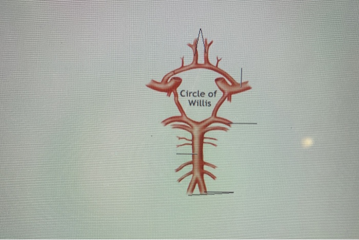
45 circle of willis without labels
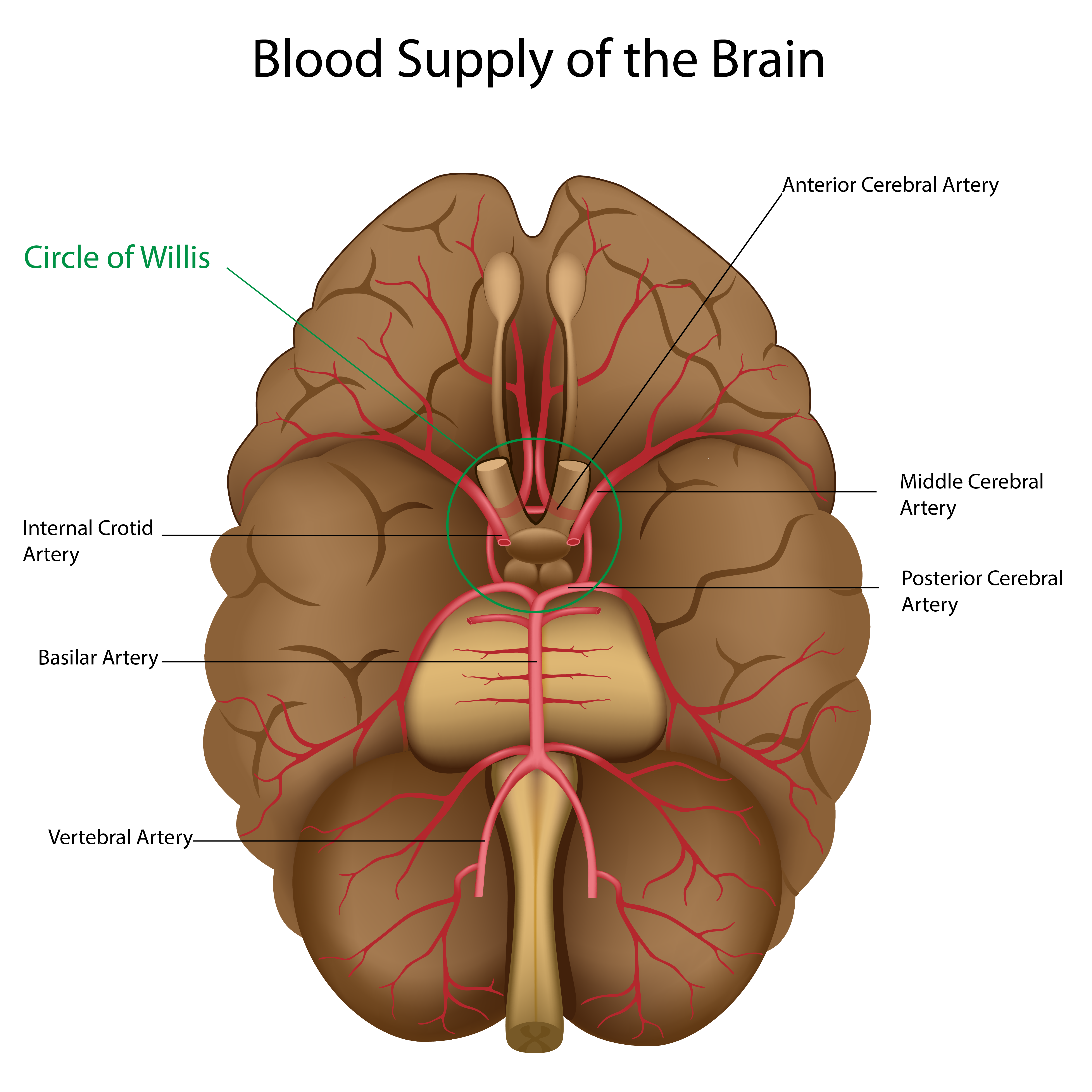
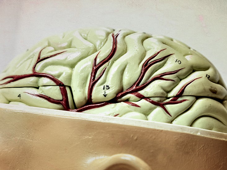
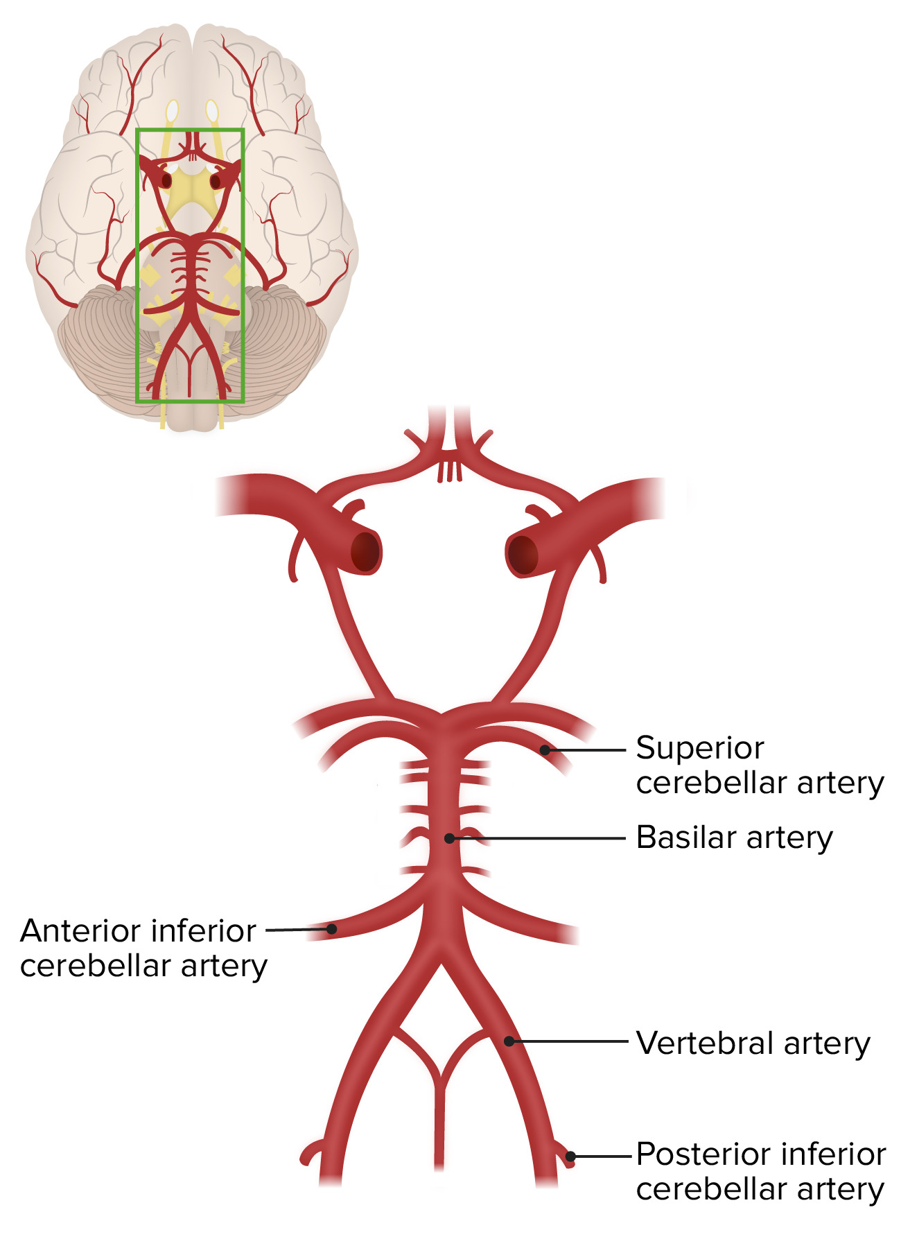
(PDF) Anatomical Labeling of the Circle of Willis Using Maximum A ... Anatomical labeling of the cerebral arteries forming the Circle of Willis (CoW) enables inter-subject comparison, which is required for geometric characterization and discovering risk factors... Cerebral Arterial Circle (Circle of Willis) | Neuroanatomy | The ... The brain has been removed to demonstrate an intact cerebral arterial circle that is formed by anastomotic connections between the basilar and internal carotid arteries. The basilar artery, formed by the union of the right and left vertebral arteries, terminates with the posterior cerebral arteries. These vessels connect to the internal carotid ...
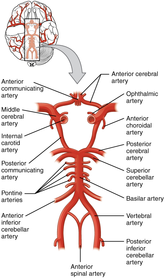
OLI - Drawing Circle of Willis - English labels | AnatomyTOOL Circle of Willis. This inferior view shows the arteries of the brain. This circular structure with its communicating branches is called the cerebral arterial circle (circulus arteriosus cerebri) or circle of Willis. English labels. Retrieved from Anatomy & Physiology by Open Learning Initiative (CC BY-NC-SA).
Circle of willis without labels
Anatomical labeling of the Circle of Willis using maximum a ... - PubMed Abstract. Anatomical labeling of the cerebral arteries forming the Circle of Willis (CoW) enables inter-subject comparison, which is required for geometric characterization and discovering risk factors associated with cerebrovascular pathologies. We present a method for automated anatomical labeling of the CoW by detecting its main bifurcations. Circle of Willis | Radiology Reference Article | Radiopaedia.org A complete circle of Willis (in which no component is absent or hypoplastic) is only seen in 20-25% of individuals. Posterior circulation anomalies are more common than anterior circulation variants and are seen in nearly 50% of anatomical specimens. Common variants hypoplasia of one or both PCOM ~30% (range 25-34%) Circle of Willis - 3D Models, Video Tutorials & Notes | AnatomyZone The Circle of Willis provides the blood supply to the brain, and essentially what happens is that it connects two arterial sources together to form this arterial circle, which then supplies the brain with blood.
Circle of willis without labels. Step 1 First Aid - Neurology Flashcards | Quizlet USMLE Step 1 First Aid - Neurology notes Learn with flashcards, games, and more — for free. Circulatory System | Circle of Willis Circulation - YouTube Official Ninja Nerd Website: Nerds!In this lecture Professor Zach Murphy will be presenting on the arterial supply to the brain, ... CT head sagittal - labeling questions | Radiology Case - Radiopaedia Normal CT head (without labels) ct Loading Image 1 CT Sagittal non-contrast The same normal brain CT without labels for reference. Case Discussion The labeled structures are (excluding the correct side): mastoid air cells temporalis muscle zygomatic arch external auditory (acoustic) canal temporomandibular joint tentorium cerebelli sylvian fissure Circle of Willis quizzes and unlabeled diagrams | Kenhub The circle of Willis, also known as the cerebral arterial circle, is formed by anterior and posterior arterial pathways. The arteries of the circle of Willis include: One anterior communicating artery Two anterior cerebral arteries (left and right) Two internal carotid arteries (left and right) Two posterior communicating arteries (left and right)
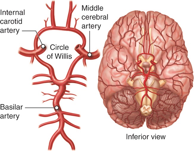
Plasticity of the adult circle of Willis in response to flow diversion ... Several studies have analyzed the artery adjacent to the device.[1, 2, 6, 10, 12, 14] A few other studies have analyzed responses in the circle of Willis and other vessels in response to flow diverter placement.[5, 7-9, 16] The phenomena reported here in the response of hypoplastic arteries make up one of the three possible types of vascular ... Circle of Willis: Anatomy, Function, and Significance - Verywell Health The circle of Willis is a group of blood vessels in the brain that connect with each other, forming a continuous structure that resembles a circle. These nine arteries supply blood to a large portion of the brain. Most of the time, blood can flow through the vessels of the circle of Willis without any interruption. What is a circle of Willis angiogram? - Studybuff The circle of Willis is a group of blood vessels in the brain that connect with each other, forming a continuous structure that resembles a circle. These nine arteries supply blood to a large portion of the brain. Most of the time, blood can flow through the vessels of the circle of Willis without any interruption. Circle of Willis - an overview | ScienceDirect Topics The circle of Willis, or the circulus arteriosus, is formed by the anastomosis of the two internal carotid arteries with the two vertebral arteries. The anterior communicating, anterior cerebral, internal carotid, posterior communicating, posterior cerebral, and basilar arteries are all part of the circle of Willis (see Fig. 3-13 ).
w-radiology.com › mra-circle-willisMRA of the Circle of Willis - W-Radiology The circle of Willis plays a crucial part in maintaining blood flow in the brain. The circle of Willis is where several arteries in the brain meet or join together(1). Also known as the circulus arteriosus cerebri or the cerebral arterial circle, the circle of Willis is an anastomotic (connecting) ring of arteries found at the base of the brain(2). This arterial circle allows the two major arterial systems in the brain, the internal carotid arteries and vertebrobasilar systems (vertebral and ... Circle Of Willis: Anatomy, Diagram And Functions - Science ABC The Circle of Willis (often abbreviated as CW or CoW) gets its name from Thomas Willis, an eminent English physician, who described the arterial ring present at the base of the brain 400 years ago. Willis wasn't the first to describe this ring of blood vessels. CIRCLE OF WILLIS - YouTube CIRCLE OF WILLIS emerson24 753 subscribers Subscribe 720 Share Save 91K views 11 years ago Easy way to remember the Circle of Willis. If you are watching this video, you most likely have a TON... Neuro Labeling - Circle of Willis, Neuro test 2 Diagram | Quizlet Connects the middle cerebral and posterior cerebral arteries. Middle cerebral artery. Supplies the lateral surface of the brain and also the deepest portions of the frontal and parietal lobes. Internal carotid artery. Supplies blood to the four lobes of the brain as well as the optic nerves and the retina of the eyes.
What is the Willis area in the brain? - Quora
Circle of Willis - 3D Models, Video Tutorials & Notes | AnatomyZone The Circle of Willis provides the blood supply to the brain, and essentially what happens is that it connects two arterial sources together to form this arterial circle, which then supplies the brain with blood.
Blood Flow Through the Circle of Willis | Ansys Courses
Circle of Willis | Radiology Reference Article | Radiopaedia.org A complete circle of Willis (in which no component is absent or hypoplastic) is only seen in 20-25% of individuals. Posterior circulation anomalies are more common than anterior circulation variants and are seen in nearly 50% of anatomical specimens. Common variants hypoplasia of one or both PCOM ~30% (range 25-34%)

Circle of Willis - 3D Anatomy Tutorial
Anatomical labeling of the Circle of Willis using maximum a ... - PubMed Abstract. Anatomical labeling of the cerebral arteries forming the Circle of Willis (CoW) enables inter-subject comparison, which is required for geometric characterization and discovering risk factors associated with cerebrovascular pathologies. We present a method for automated anatomical labeling of the CoW by detecting its main bifurcations.

0514 Cerebral Arterial Circle Of Willis Medical Images For ...

Circle of Willis: Anatomy, function, and what to know

Brain arteriovenous malformations: A scoping review of ...
Circle of Willis: Anatomy and function | Kenhub
Posterior Cerebral Artery Anatomy, Function & Diagram | Body Maps

The 17 Circle of Willis variants used in the analyses. "N ...

Circle of Willis - Wikipedia

Circle Willis Stock Vector (Royalty Free) 24963922 | Shutterstock
Circle Of Willis: Anatomy, Diagram And Functions » Science ABC

Circle of Willis - an overview | ScienceDirect Topics
Solved 9. Label the following arteries: anterior cerebral ...
Circle of Willis: Anatomy, function, and what to know

Aaron Rutman, MD on Twitter: "3/3 ...while others use the ...

Anatomy of the aorta: location and branches | GetBodySmart
OLI - Drawing Circle of Willis - English labels | AnatomyTOOL

Acetylcholine - The first neurotransmitter ever discovered ...
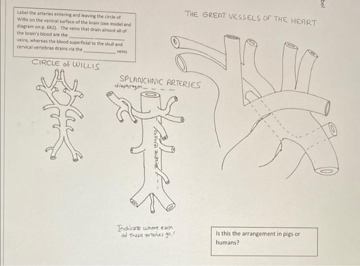
Solved Color and label the veins (p. 942) and arteries (p ...

Radiopaedia - Drawing Circle of Willis - no labels | AnatomyTOOL

Circle of Willis - an overview | ScienceDirect Topics

ImageQuiz: Circle of Willis | Label

Neuro Labeling - Circle of Willis Diagram | Quizlet

Circle of Willis | Radiology Reference Article | Radiopaedia.org

Circle of Willis - JLC Flashcards | Quizlet

24 Angio Mri Photos and Premium High Res Pictures - Getty Images
:max_bytes(150000):strip_icc()/CircleofWillis-87378170-3ece0502a02949dd82310d723e0d4c98.jpg)
Circle of Willis: Anatomy, Function, and Significance
Blood supply of the brain

Circle of Willis Label Diagram | Quizlet

9 Best Circle of Willis ideas | circle of willis, circle ...
Solved] Draw the Circle of Willis, and label the vessels ...

Circle Christmas Sheet Labels – Label-Headquarters.com
Circle of Willis: Anatomy and function | Kenhub
Computational analysis of renal artery flow characteristics ...
Cerebrovascular System: Anatomy | Concise Medical Knowledge

Circle of Willis (Without Aneurysms) | United Biologics

0514 Cerebral Arterial Circle Of Willis Medical Images For ...

Circle Willis Stock Illustration 200175368 | Shutterstock

Circle of Willis labeling Diagram | Quizlet

Circle of Willis | Radiology Reference Article | Radiopaedia.org
OpenStax AnatPhys fig.13.15 - Circle of Willis - English ...

Circle of Willis: Anatomy, function, and what to know

The Arterial Circle Of Willis Quiz - ProProfs Quiz

File:Circle of Willis unlabeled.svg - Wikimedia Commons
Post a Comment for "45 circle of willis without labels"