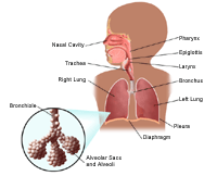
44 label the photomicrogram of the lung
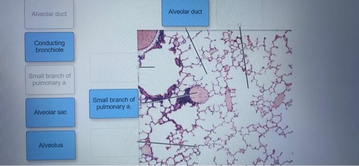
Solved Label the photomicrogram of the lung Alveolar duct ... Label the photomicrogram of the lung Alveolar duct Alveolus Small branch of pulmonary a. bronchiole Alveolar sac Reset Zoom < Prev 22 of 40::: Next > 20 This problem has been solved! You'll get a detailed solution from a subject matter expert that helps you learn core concepts. See Answer Respiratory System | histology - University of Michigan For descriptive purposes the respiratory system is divided into (a) conducting and (b) respiratory portions. The conducting part consists of passageways which carry air to the respiratory portion. The respiratory portion begins at the level where alveoli first appear in the final branches of the bronchioles.
A&P 2 Lab Respiratory System Flashcards | Quizlet Label the photomicrogram of the lung. Identify the cartilaginous anatomical structures shown in the posterior view of the superior portion of the lower respiratory system. Place the following structures with the appropriate anatomical region.

Label the photomicrogram of the lung
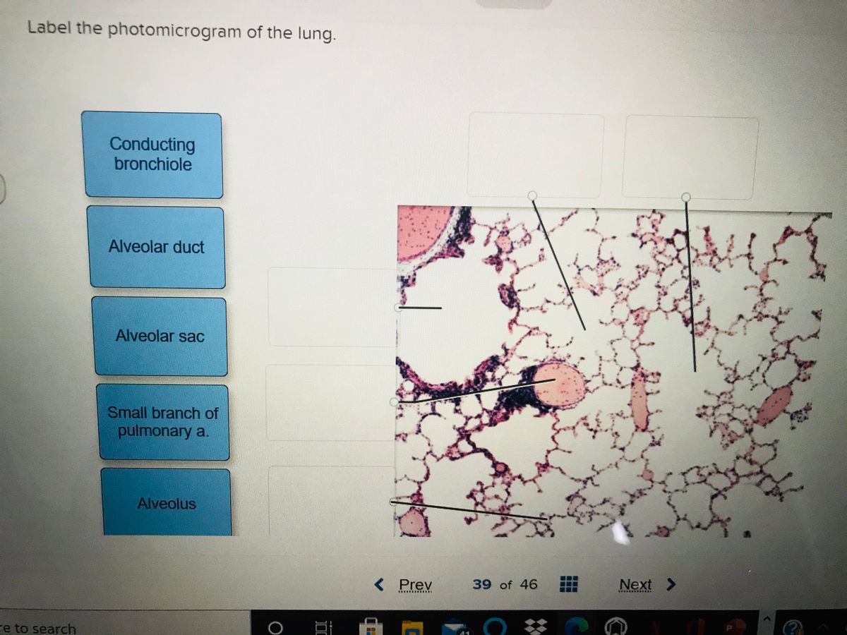
Answered: Which structure is highlighted? | bartleby WebTranscribed Image Text: Label the photomicrogram of the lung. Conducting bronchiole Alveolar duct Alveolar sac Small branch of pulmonary a. Alveolus < Prev 39 of 46 # Next … Solved Label the photomicrogram of the lung … WebLabel the photomicrogram of the lung Segmental branch of pulmonary a. Interalveolar wall Alveolar macrophage Reset Zoom < Prev 19 40 , Next > 2 3 4 5 6 This problem has been solved! You'll get a detailed solution from … A photomicrograph of (a) group I lung tissues showing … WebA photomicrograph of (a) group I lung tissues showing normal alveolar... | Download Scientific Diagram A photomicrograph of (a) group I lung tissues showing normal alveolar sacs (AS), alveoli...
Label the photomicrogram of the lung. BIOL 320 - Practical 2 - Lab 7 Flashcards | Quizlet WebStudy with Quizlet and memorize flashcards containing terms like Label the photomicrograph using the hints provided., Label the types of cells in the … A&P 139 Chapter 19 Flashcards | Quizlet WebView the illustration, and drag the labels to their appropriate place. pressure in alveoli increases and their walls burst, coalescing them and decreasing the surface area for gas … photomicrograph of lung sections WebFigure 1: A photomicrograph of lung sections from (a) a control group showing normal epithelization of bronchi and bronchioles with normal alveoli and normal configuration of … A&P 2 Lab Respiratory System Flashcards | Quizlet WebLabel the photomicrogram of the lung. Identify the cartilaginous anatomical structures shown in the posterior view of the superior portion of the lower respiratory system. Place …
Bronchi: Anatomy, function and histology | Kenhub Oct 25, 2022 · Bronchi. Bronchi are plural for bronchus and represent the passageways leading into the lungs. The first bronchi branch from trachea, and they are the right and left main bronchi. These bronchi are the widest and they enter the lung. After entering the lungs, the bronchi continue to branch further into the secondary bronchi, known as lobar ... Solved Alveolar duct histology respiratory system Label the ... Alveolar duct histology respiratory system Label the photomicrogram of the lung. This problem has been solved! You'll get a detailed solution from a subject matter expert that helps you learn core concepts. See Answer Question: Alveolar duct histology respiratory system Label the photomicrogram of the lung. Show transcribed image text Expert Answer Lung | Radiology Reference Article | Radiopaedia.org Mar 27, 2023 · The lungs are the functional units of respiration and are key to survival. They contain 1500 miles of airways, 300-500 million alveoli and have a combined surface area of 70 square meters (half a tennis court). Each lung weighs approximately 1.1 kg. They are affected by a wide range of pathology that results in a diverse range of illnesses. A&P 2 Lab Unit 2 Flashcards | Quizlet Label the photomicrogram of the lung. Identify the cartilaginous anatomical structures shown in the posterior view of the superior portion of the lower respiratory system. Place the following structures with the appropriate anatomical region. Labels maybe be placed in more than one category.
Cambridge International Examinations Cambridge International … Web1 Fig. 1.1 is a photomicrograph of epithelial cells in the bronchus. $ ... Smoking causes damage to the epithelial cells of the lungs. ... Draw a label line and label on Fig. 4.1 to … Solved Label the photomicrogram of the lung Alveolar duct - Chegg WebLabel the photomicrogram of the lung Alveolar duct Alveolus Small branch of pulmonary a. bronchiole Alveolar sac Reset Zoom < Prev 22 of 40::: Next > 20 This problem has … A photomicrograph of (a) group I lung tissues showing … WebA photomicrograph of (a) group I lung tissues showing normal alveolar... | Download Scientific Diagram A photomicrograph of (a) group I lung tissues showing normal alveolar sacs (AS), alveoli... Solved Label the photomicrogram of the lung … WebLabel the photomicrogram of the lung Segmental branch of pulmonary a. Interalveolar wall Alveolar macrophage Reset Zoom < Prev 19 40 , Next > 2 3 4 5 6 This problem has been solved! You'll get a detailed solution from …
PDF) Blunted Activation of Rho-Kinase in Yak Pulmonary ...
Answered: Which structure is highlighted? | bartleby WebTranscribed Image Text: Label the photomicrogram of the lung. Conducting bronchiole Alveolar duct Alveolar sac Small branch of pulmonary a. Alveolus < Prev 39 of 46 # Next …
Research Article Prolonged Inhalation Exposure to Coal Dust ...
Pleural Fluid" Images – Browse 716 Stock Photos, Vectors, and ...
JaypeeDigital | eBook Reader
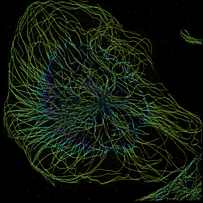
3D Nanoscale architecture of microtubule network | 2017 ...
Solved General Structure of Mucosa Label the structures that ...

a, b): Photomicrographs of sections of control rat lung ...

CHoMP: A Chemoenzymatic Histology Method Using Clickable ...
Anatomy of the Respiratory System in Children

Chol Mayar Anai (cholmayar16) - Profile | Pinterest

Curcumin and its nano-formulation: The kinetics of tissue ...

A&P 2 Lab Practical Final Flashcards | Quizlet
3D Nanoscale architecture of microtubule network | 2017 ...
Review Article Robotic and Microrobotic Tools for Dental Therapy

The Cyclin-dependent Kinase Inhibitors p15INK4B and p21CIP1 ...

Target Inhibition of IL-1 Receptor Prevents Ifosfamide ...

The Pathologist September 2020 by texerepublishing - Issuu
Test Code Test Name Components Specimen Method Comments ...
Untitled
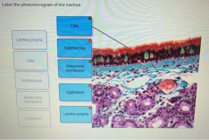
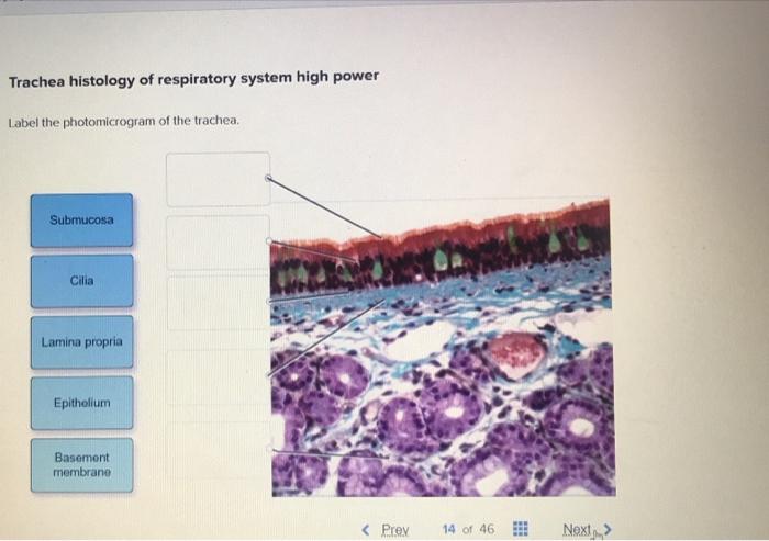
Solved Label the photomicrogram of the trachea. Cilia Lamina ...

Prolonged Inhalation Exposure to Coal Dust on Irradiated Rats ...

BIR Publications
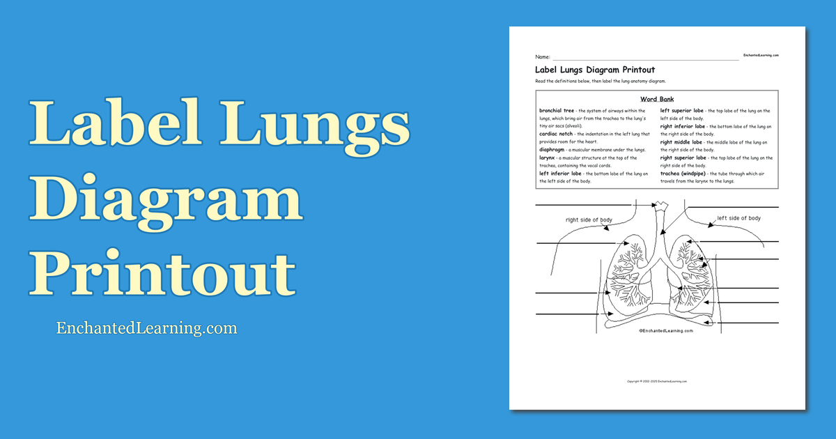
Label Lungs Diagram Printout - Enchanted Learning
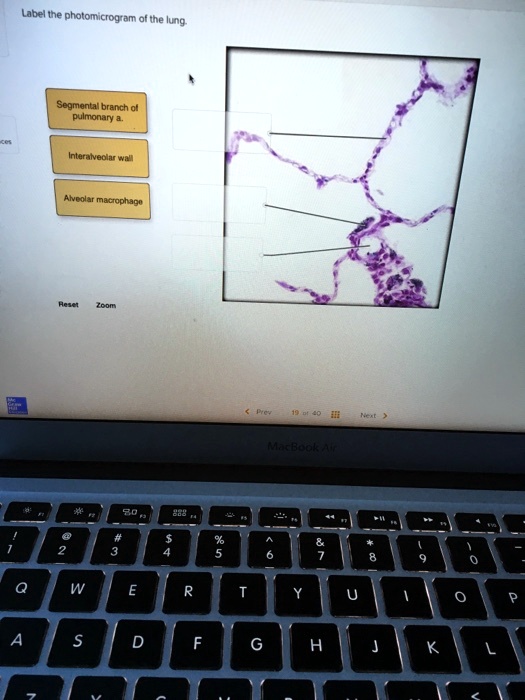
SOLVED: Label the photomicrogram of the lung Segmental branch ...
Solved Label the photomicrogram of the lungcan be answered ...
clover/keywords.txt at master · epireve/clover · GitHub
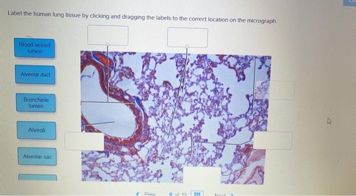
Solved Label the human lung tissue by clicking and dragging ...
Answered: Which structure is highlighted? | bartleby
Usage of pharmaceutical contraceptive drug for controlling ...
pp97-144 Vol 59(3) FINAL.pmd
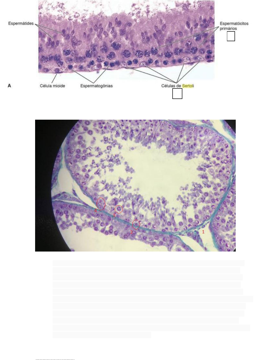
SISTEMA REPRODUTOR MASCULINO - Histologia I
fenrg-10-922169-g006.jpg

Biology 234 ~ Lab MIDTERM Practical Flashcards | Quizlet
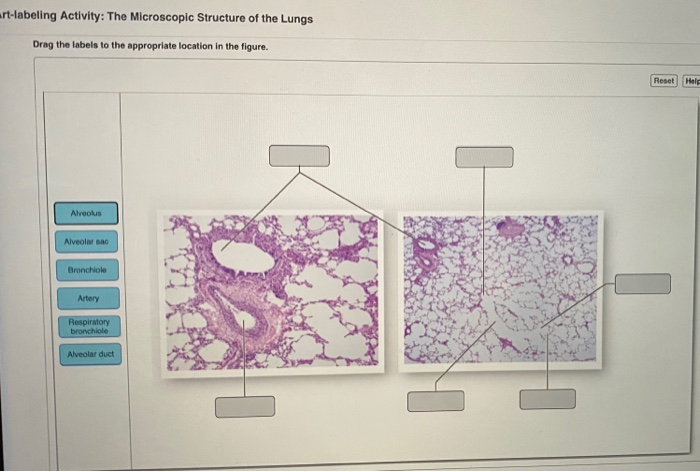
Solved art-labeling Activity: The Microscopic Structure of ...
BreadAndButter/space-dict.txt at master · DreamSea ...
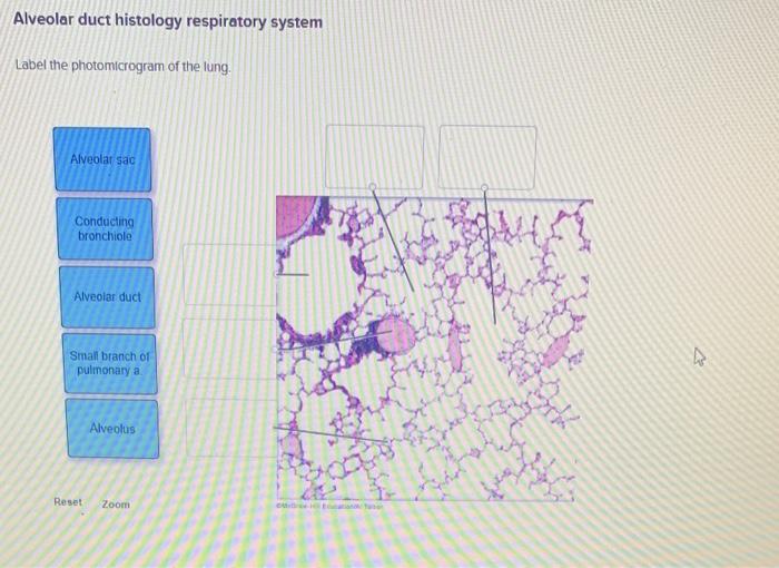
Solved Alveolar duct histology respiratory system Label the ...
Scheme of Examination o o
Medicina | May 2021 - Browse Articles

UNIT 1 HW & Quizzes Flashcards | Quizlet

A&P 2 Lab Unit 2 Flashcards | Quizlet
Kidney-Targeted Naked DNA Transfer by Retrograde Renal Vein ...
Abstracts of Twenty-Sixth Annual Meeting of the Radiation ...
Commission
Popular Electronics' ELECTRONIC
Post a Comment for "44 label the photomicrogram of the lung"